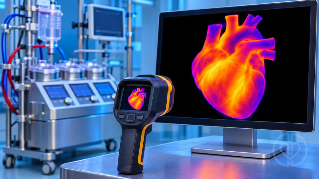
Infrared thermography may reveal clinically relevant cooling that a systemic temperature reading misses during cardiopulmonary bypass (CPB). In a five-patient retrospective case series, Nishimura and colleagues examined its use during valve surgery in patients with cold agglutinin disease. Their observations draw attention to two vulnerable locations: the exposed epicardial surface and the cardioplegia delivery line.
Why temperature location matters
Cold-reactive antibodies can cause red blood cell agglutination and complement-mediated hemolysis. The thermal amplitude—the highest temperature at which agglutination occurs—is therefore a central consideration when planning bypass and myocardial protection. A satisfactory systemic temperature does not establish that every exposed surface or fluid pathway remains above that threshold.
This report explores a practical monitoring adjunct rather than a replacement for conventional temperature monitoring or individualized hematology and surgical planning.
Study design and temperature strategy
The investigators identified five patients with cold agglutinin disease among 416 open-heart procedures performed between September 2013 and December 2023. All five underwent elective valve surgery. One early patient was managed without thermography; the next four received thermographic monitoring alongside a broader warming and myocardial-protection strategy.
Systemic CPB temperatures were maintained at 34–36°C, with intended cardioplegia temperatures of 28–32°C. The operating room was kept warm, and intravenous medications and blood products were prewarmed. These were the institution’s case-specific management choices, not validated temperature targets for every patient with cold-reactive antibodies.
A compact infrared camera was positioned approximately 80 cm above the surgical field. The team sampled three epicardial points and averaged their temperatures. When surface cooling approached an individual patient’s thermal amplitude, warm saline irrigation was used and the temperature response was checked. Cardioplegia-line measurements were compared with a circuit thermometer, with recalibration when discrepancies appeared.
What infrared thermography detected
Epicardial cooling despite warmer systemic temperatures
In the four monitored patients, the epicardial surface was an average of 3.1°C cooler than the target systemic temperature, with differences ranging from 1.7 to 4.1°C. This finding illustrates how exposure after sternotomy can create a local temperature gap even when systemic management appears satisfactory.
Cool residual prime in the cardioplegia line
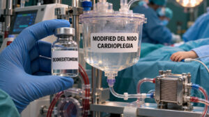
Before drainage of residual priming solution, reported cardioplegia-line temperatures averaged 23.3°C, ranging from 21.5 to 25.9°C. These readings were below the intended cardioplegia range. The reported average 11.4°C difference was relative to the target systemic temperature; it should not be interpreted as a measured 11.4°C fall across the cardioplegia heat exchanger.
The team drained residual prime until the intended delivery temperature was confirmed before administering cardioplegia. The observation reinforces the distinction between a temperature setting and the temperature actually present near delivery.
Clinical outcomes and limits of interpretation
All five patients survived to discharge. The four thermography-monitored patients were described as having favorable postoperative courses. The earlier unmonitored patient developed myocardial injury, a decline in ejection fraction from 69% to 39%, and a rhythm disturbance requiring permanent pacing.
That contrast does not prove that thermography prevented myocardial injury. The groups comprised only four patients and one historical patient, without randomization or a comparable control group. The unmonitored case also involved a longer cross-clamp time and a different operative context. Cold-related microcirculatory injury was a proposed explanation, not a directly demonstrated mechanism; myocardial stunning, cardioplegia distribution, embolic injury, and other causes remained possible.
The paper also contains a reporting inconsistency: the abstract gives a thermal-amplitude range of 19–26°C, whereas Table 1 and the main results include a patient at 15°C. Thermography measures surface radiation rather than deep myocardial or coronary microcirculatory temperature, and readings can be affected by reflections and ambient conditions.
Relevance for perfusion practice
The most useful message is the need to distinguish systemic, epicardial, and cardioplegia-delivery temperatures. Infrared thermography may provide additional spatial information during sternotomy, bypass initiation, and cardioplegia delivery. This preliminary experience supports further evaluation of that role, while leaving its effect on clinical outcomes unresolved.
Source
Nishimura Y, Kuwahara G, Wada H. Feasibility and Utility of Infrared Thermography for Intraoperative Temperature Monitoring in Cold Agglutinin Disease during Cardiopulmonary Bypass. Annals of Thoracic and Cardiovascular Surgery. 2026;32:26-00158. doi:10.5761/atcs.oa.26-00158.
Disclosures: The authors reported no specific research funding and no competing interests. This educational summary discusses the uploaded publication and does not establish a clinical protocol.