
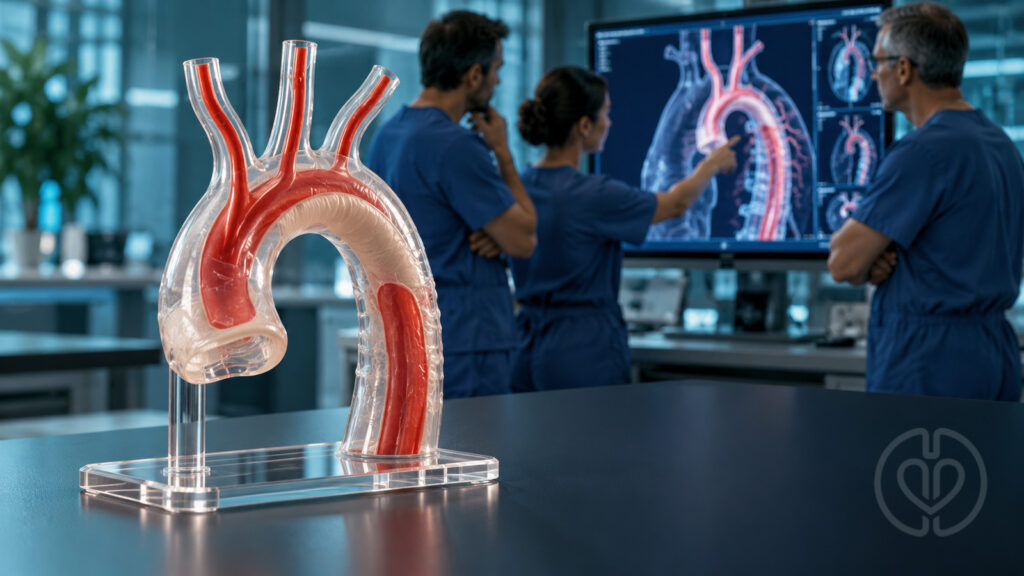
Multidisciplinary collaboration in aortic surgery requires more than having the right specialists in the operating room. A new American Heart Association scientific statement explains how decisions by the surgeon, anesthesiologist, and perfusionist immediately affect one another during complex aortic repair. DeAnda and colleagues examine acute type A aortic dissection and open thoracic or thoracoabdominal aortic surgery, emphasizing shared planning and real-time adjustment as circulation, ventilation, and organ-protection requirements change.
Why Aortic Surgery Demands Continuous Coordination
The statement complements existing aortic disease guidance rather than providing an exhaustive operative manual. Its central concern is the interaction among specialties: a change in cannulation can alter pressure-monitor interpretation, lung isolation can change oxygenation, and extracorporeal drainage can reduce the native cardiac output needed to perfuse the upper body.
The multidisciplinary aortic team also includes nurses, physician assistants, surgical technologists, and other clinicians. The document concentrates on three core intraoperative viewpoints to illustrate why technically sound individual actions must fit a coordinated plan.
Acute Type A Dissection: Connecting Repair and Perfusion Plans
For acute type A dissection, the extent of repair depends on the entry tear, root involvement, arch anatomy, malperfusion, and patient stability. The statement describes a tear-oriented strategy and an individualized choice between limited and more extensive reconstruction. A longer or more complex repair can change the anticipated duration of circulatory arrest and the cerebral-protection plan.
Arterial access is therefore a joint surgical and perfusion decision. Axillary or innominate cannulation can facilitate antegrade cerebral perfusion, whereas femoral access may provide rapid support but requires attention to retrograde perfusion and malperfusion. Direct aortic cannulation is another option when the true lumen can be safely accessed and confirmed with imaging. No single access strategy is presented as appropriate for every dissection.
Cerebral Protection During Circulatory Arrest
Hypothermia reduces cerebral metabolic demand, but temperature alone does not remove the risks of interrupted circulation. The statement discusses antegrade cerebral perfusion (ACP) and retrograde cerebral perfusion (RCP) as adjuncts, with the choice influenced by anatomy, cannulation, expected arrest duration, and institutional experience.
ACP supplies blood through the arterial circulation and can support cerebral protection during more prolonged reconstruction. RCP delivers blood through the superior vena cava and is commonly combined with deeper cooling for shorter arrest periods. The authors review evidence supporting both approaches while recognizing continuing debate over the optimal strategy. The summary should not be interpreted as proof that one method is universally superior.
Cooling, initiation of selective cerebral perfusion, cessation of systemic flow, reperfusion, and rewarming are critical communication points. The entire team needs to understand which territories are receiving blood, which pressure measurements remain meaningful, and whether the operative plan has changed. The paper does not establish one universally applicable temperature or flow prescription.
Monitoring Works Best When the Team Shares Its Interpretation
Pressure monitoring and echocardiography
Arterial pressure readings depend on the relationship between monitoring sites, cannulas, clamps, and the operative field. A radial measurement that is useful during one phase may become interrupted or misleading during another. The statement discusses multiple-site pressure monitoring and transesophageal echocardiography to assess true-lumen access, cardiac function, drainage, and ventricular filling.
Cerebral oxygenation and electrical activity
Near-infrared spectroscopy (NIRS) provides regional cerebral oxygenation trends, while electroencephalographic monitoring offers information about cerebral electrical activity and anesthetic effects. Neither represents a complete measurement of brain perfusion or guarantees freedom from neurologic injury.
A falling NIRS value can reflect cannula position, inadequate flow or pressure, impaired oxygen content, ventilation-related carbon dioxide changes, or other patient and circuit factors. The statement uses this problem to illustrate coordinated troubleshooting: anesthesia, surgery, and perfusion may need to investigate several causes at the same time. Interpretation must account for the monitored region and physiologic confounders.
Thoracoabdominal Repair: Protecting the Spinal Cord and Distal Organs
Cross-clamping the descending or thoracoabdominal aorta can produce proximal hypertension and distal ischemia. Preoperative assessment therefore includes the extent of disease and cardiac, pulmonary, renal, and neurologic risk. In elective cases, the statement discusses opportunities to address anemia, smoking, functional capacity, and other modifiable risks before surgery.
Spinal cord protection can include cerebrospinal fluid drainage, maintenance of perfusion pressure, and selected motor or somatosensory evoked-potential monitoring. The effectiveness and interpretation of neuromonitoring also depend on anesthetic and neuromuscular-blocking agents. Drain management and responses to signal changes require a defined multidisciplinary plan rather than isolated adjustments by one clinician.
Choosing and Managing Distal Perfusion
The statement reviews several approaches, including clamp-and-sew, passive shunts, left heart bypass, conventional cardiopulmonary bypass (CPB), and extracorporeal membrane oxygenation (ECMO). These options differ in drainage, oxygenation capability, anticoagulation requirements, temperature control, and handling of shed blood.
| Approach | Key coordination issue |
|---|---|
| Left heart bypass | Returns blood drained from the left atrium to the distal arterial circulation; native cardiac output and lung function remain important. |
| CPB | Provides extracorporeal oxygenation and systemic support, with cardiotomy suction available and full anticoagulation required. |
| ECMO used for operative support | Provides extracorporeal oxygenation through a closed circuit; configuration and anticoagulation must fit the operation. |
During partial support, excessive drainage can reduce ventricular filling and jeopardize native output to the coronary and cerebral circulations. Insufficient drainage may limit distal support. Echocardiography and pressure monitoring help anesthesia and perfusion balance these competing needs while the surgeon manages the clamped aorta.
Lung Isolation and Changing Support Requirements
Open descending and thoracoabdominal exposure often requires right-lung ventilation with isolation of the left lung. Airway selection depends on anatomy, compression, access, and postoperative ventilation needs. The authors discuss the tradeoffs between a double-lumen tube and a bronchial blocker without identifying a universally preferred device for thoracoabdominal surgery.
The relative contributions of native lungs and extracorporeal oxygenation can change during the procedure. Communicating those transitions helps the team interpret oxygenation and ventilation problems, particularly when partial bypass still depends on blood passing through the ventilated lung.
Evidence Limits and Practical Implications
This is a scientific statement synthesizing existing literature and expert perspectives, not a new randomized trial of teamwork or perfusion technique. The authors identify gaps in comparative evidence for cerebral and spinal cord protection, monitoring, and perfusion management. Pharmacologic suppression of cerebral metabolism also does not reliably establish improved neurologic outcomes.
For perfusion practice, the most useful message is to make transitions explicit: confirm the operative plan, anticipated arrest and perfusion phases, meaningful monitoring sites, oxygenation responsibilities, and responses to evolving conditions. The statement supports multidisciplinary training and simulation, while calling for prospective multicenter studies to define best practices.
The source includes writing-group and reviewer disclosure tables. These should be consulted for individual relationships; the statement should not be described as free of disclosures. The uploaded publication retains provisional page and publication-date placeholders, so the DOI is the most precise citation identifier available in this copy.
Original Article
DeAnda A Jr, Steppan J, Boskovski MT, et al. Multidisciplinary collaboration in the performance of aortic surgery: a scientific statement from the American Heart Association. Circulation. 2026;154. doi:10.1161/CIR.0000000000001490.
This independently written educational summary accompanies the unchanged source PDF. Source copyright belongs to the American Heart Association. The cover is an AI-generated editorial illustration.