Ascending aortic calcium may reveal cardiovascular risk that remains hidden when myocardial perfusion, myocardial flow reserve, and coronary calcium are all normal. In a 13-site REFINE-PET registry analysis, automated measurement of ascending aortic calcification was associated with subsequent cardiovascular events, particularly heart failure admission. The findings suggest a potentially useful role for information already present on routine PET/CT images.
Preprint notice: This report was posted on medRxiv on October 12, 2026, and has not been certified by peer review. Its findings are preliminary and should not independently guide clinical practice.
Study Design and the Triple-Normal Population
Spielvogel and colleagues retrospectively analyzed clinically acquired myocardial perfusion PET/CT examinations from 13 centers between 2007 and 2020. The full eligible cohort included 21,923 patients. The primary analysis focused on 3,580 patients, representing 16.3% of the cohort, who met all three normal imaging criteria: stress total perfusion deficit below 5%, myocardial flow reserve above 2, and a coronary artery calcium score of zero.
This distinction matters because a normal coronary and myocardial evaluation does not directly characterize the entire arterial system. The study asked whether calcification in the ascending aortic wall could identify residual risk within a population otherwise considered reassuring by these imaging measures. Median age was 60 years, and 64.7% were women. Hypertension and diabetes remained common despite normal imaging results.
Measuring Ascending Aortic Calcium With AI
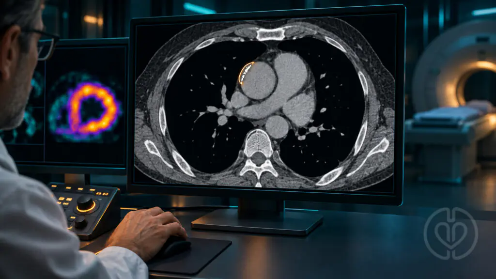
The investigators used TotalSegmentator to identify the aorta on the noncontrast, low-dose CT scans acquired for PET attenuation correction. Subsequent image processing separated the ascending and descending thoracic aorta, isolated a wall region, excluded adjacent bone, and identified candidate calcium at a threshold of at least 130 Hounsfield units. Small lesions were removed to reduce noise, and segment-specific Agatston scores were calculated.
The approach uses existing CT data, so calcium measurement itself requires no additional image acquisition, contrast administration, or radiation exposure. That does not establish that implementation, software integration, quality assurance, or follow-up care would have no cost. The analysis required suitable CT coverage and successful automated segmentation; the aortic arch could not be assessed reliably because it was frequently outside the available field of view.
A reader substudy compared automated measurements with assessments by three experienced readers in 244 scans from 122 patients. Agreement with the mean reader score was good to excellent, with intraclass correlation coefficients of 0.87 for rest acquisitions and 0.90 for stress acquisitions. Manual annotation took an average of 4.7 minutes per scan, illustrating the workload that automation might reduce.
Key Findings
Ascending aortic calcification was present in 1,322 patients, or 36.9% of the triple-normal group. Among patients with calcium, the median ascending aortic Agatston score was 65. Over a median follow-up of approximately 52 months, 194 primary composite events occurred, corresponding to 1.15 events per 100 patient-years.
| Outcome | Adjusted hazard ratio | 95% confidence interval |
|---|---|---|
| Cardiovascular death, myocardial infarction, or heart failure admission | 1.24 | 1.09–1.40 |
| Heart failure admission | 1.30 | 1.10–1.55 |
| All-cause mortality | 1.17 | 1.04–1.32 |
The continuous calcium score was transformed using log(1 + score) because many patients had zero calcium and the distribution was skewed. The hazard ratios therefore should not be interpreted as effects per single Agatston unit or per fixed raw-score increment. A separate analysis comparing any ascending aortic calcium with none found an adjusted hazard ratio of 1.44 for the primary composite endpoint, with a 95% confidence interval of 1.08–1.91.
Heart Failure Drove Much of the Signal
Among the 194 primary events, 95 were heart failure admissions, 65 were myocardial infarctions, and 34 were cardiovascular deaths without a preceding heart failure admission or myocardial infarction during follow-up. Heart failure admission showed the strongest adjusted association with ascending aortic calcium. The separate adjusted associations with myocardial infarction and cardiovascular death were not statistically significant; their confidence intervals crossed 1.
The authors propose that aortic calcium may reflect vascular stiffening and increased ventricular afterload. This is a plausible explanation for the prominence of heart failure, but the study did not directly measure aortic stiffness, pulse-wave velocity, or augmentation index. It also did not establish the subtype of heart failure responsible for the association. Preserved baseline ejection fraction alone cannot demonstrate that later admissions represented heart failure with preserved ejection fraction.
Statistical Association Versus Useful Prediction
Adding ascending aortic calcium improved model fit beyond clinical variables, including models that also contained myocardial flow reserve and stress perfusion deficit. However, the gain in discrimination was small: the reported C-statistic improvement was approximately 0.007 for the composite endpoint and 0.009 for heart failure admission. The bootstrap confidence intervals for both improvements included zero.
This distinction prevents overstating the result. A marker can remain statistically associated with outcomes while adding only limited ability to distinguish which individual patients will experience an event. The analysis supports further evaluation of opportunistic aortic calcium reporting, but does not yet establish a clinically actionable threshold or demonstrate that acting on the score improves patient outcomes.
Relevance for Perfusion and Cardiovascular Teams
For perfusionists and multidisciplinary cardiovascular teams, the report reinforces the difference between normal myocardial perfusion imaging and the broader burden of vascular disease. Here, “perfusion” refers to PET assessment of myocardial blood flow, rather than cardiopulmonary bypass performance. The study does not test cannulation strategy, aortic manipulation, embolic protection, bypass management, or surgical risk prediction.
The potential contribution is an additional imaging marker available from an existing examination. Whether it should trigger a particular heart failure assessment or preventive intervention requires prospective validation. The findings should not be extrapolated directly to operative decisions or used to replace established patient assessment.
Limitations and Disclosures
This was a retrospective observational analysis, so residual confounding remains possible. Patients with aortic calcium were older and differed in hypertension, diabetes, renal function, and other characteristics. Statistical adjustment reduces measured confounding but cannot exclude effects from unmeasured factors such as mineral metabolism, inflammation, or duration of hypertension.
Absolute event rates remained relatively low, and events also occurred in patients without ascending aortic calcium. Absence of calcium therefore did not eliminate risk. Scanner and protocol differences across sites, incomplete aortic coverage, and acquisition variability also matter. Although the authors discuss potential use in SPECT/CT and oncology PET/CT, those applications were not evaluated in this study.
The work received Austrian Science Fund and NIH support. The authors report consulting, research, speaking, software royalty, and industry relationships detailed in the source manuscript. These disclosures are relevant when considering automated imaging software and its potential clinical adoption.
Original Article
Spielvogel CP, Shanbhag A, Khanna S, et al. Ascending Aortic Calcium Identifies Patients at Risk Despite Normal Myocardial Flow, Perfusion and Coronary Calcium: A 13-Site Cohort Study. medRxiv [preprint]. Posted October 12, 2026. doi:10.64898/2026.10.09.26365170.
The source PDF is unchanged. The cover is an AI-generated editorial illustration, not a patient scan or validated diagnostic image.