AI in valvular heart disease is expanding from automated image measurements to screening, procedural planning, and outcome prediction. A state-of-the-art review by Maznyczka and colleagues examines how these tools may support Heart Teams across the care pathway. The central distinction is between technical performance and clinical benefit: a model that measures anatomy accurately or predicts risk still needs evidence that using it improves decisions and patient outcomes.
What This Review Covers
Published in JACC: Cardiovascular Interventions, the review brings together applications involving digital stethoscopes, electrocardiograms, echocardiography, computed tomography (CT), patient-specific simulation, and intraprocedural image guidance. It also discusses emerging digital twins and simulation-based training.
This is a narrative synthesis of technologies at different stages of development. It does not enroll a new patient cohort, pool treatment effects in a meta-analysis, or establish a single benefit estimate for AI-assisted valve care. Commercial tools, research algorithms, and future concepts should therefore be interpreted separately.
AI in Valvular Heart Disease: Detection and Assessment
Screening With Auscultation and Electrocardiography
Digital stethoscopes and AI-enhanced electrocardiograms may help identify patients who need further valve assessment. However, the review describes substantial variation in performance between systems and target conditions. A positive screening result requires clinical evaluation and appropriate imaging; an algorithm’s classification is not a complete valve diagnosis.
One example is ValveNet, developed and validated across multiple sites in more than 77,000 patients. The review reports 78% sensitivity, 73% specificity, and an area under the receiver-operating characteristic curve of 0.84 for detecting at least moderate aortic stenosis, aortic regurgitation, or mitral regurgitation. These results concern a particular model and diagnostic target, and should not be generalized to every AI-enabled ECG system.
Echocardiography and CT
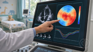
Echocardiographic applications include guiding image acquisition, identifying standard views, tracing anatomical structures, and grading valve dysfunction. Automated analysis may reduce repetitive work and help clinicians recognize disease patterns. Image quality remains a major constraint: performance on carefully selected training images may deteriorate when a model encounters suboptimal studies in routine practice.
CT applications include identifying valve calcification and segmenting the aortic root, annulus, and related structures. These capabilities can support disease assessment and structural heart planning, but automated measurements still require review for anatomical plausibility and suitability for the intended procedure.
Planning Valve Intervention
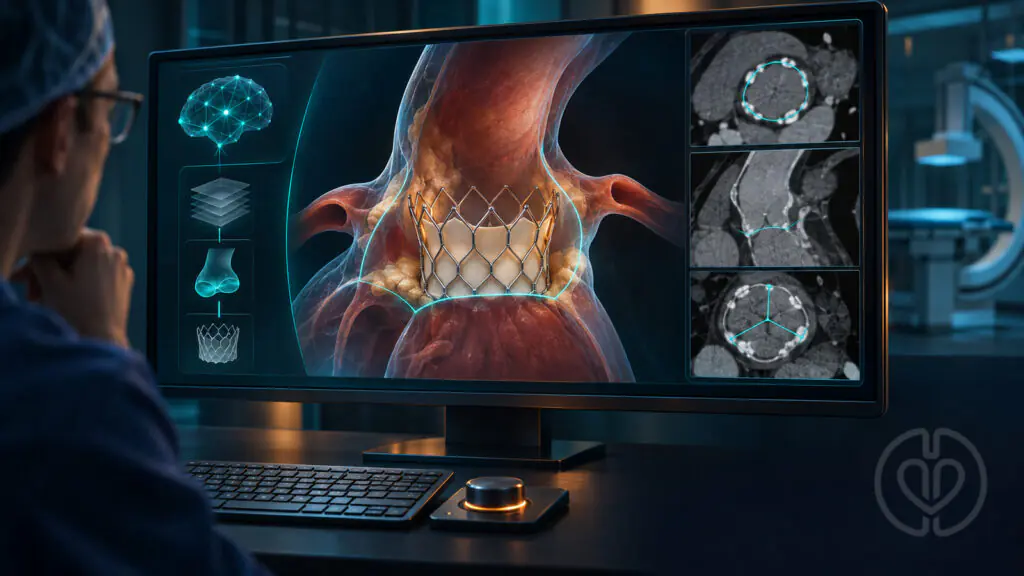
For transcatheter aortic valve replacement (TAVR), automated CT analysis can help characterize annular dimensions, access anatomy, and relevant landmarks. The review distinguishes geometric measurements from patient-specific simulations that also model interactions between a prosthesis and surrounding tissue.
These simulations may estimate frame expansion, paravalvular leak, coronary obstruction, or other complications under different implant scenarios. Their outputs depend on the underlying anatomy, device model, and mechanical assumptions. Validation against postprocedural imaging is valuable, but it does not by itself establish that simulation-guided planning improves clinical outcomes.
Mitral and tricuspid applications include automated annular assessment and virtual valve placement. In transcatheter mitral valve replacement, prediction of the residual left ventricular outflow tract—the neo-LVOT—can help assess obstruction risk. The authors caution that simulations assuming uniform valve expansion may miss important patient-specific tissue and device interactions. Simulations for mitral transcatheter edge-to-edge repair remain largely exploratory.
Guidance During the Procedure
Image fusion combines complementary information from echocardiography, fluoroscopy, and sometimes CT. A shared anatomical view can help the imager and operator communicate about device position, orientation, and the intended target. Emerging systems also automate device tracking and the reconstruction of useful imaging planes.
The underlying images and their alignment still matter. Preprocedural CT represents a static dataset, and inaccurate registration can misalign the overlay with the patient’s anatomy. Better visualization is a useful technical advance, while claims of fewer complications or improved survival require direct clinical evaluation.
Risk Prediction and the Digital Twin Concept
Machine learning can combine clinical findings, imaging, frailty measures, and procedural information to estimate risk. Some models discussed in the review performed better than conventional scores in their study populations. The authors also emphasize that AI does not consistently outperform traditional statistical methods, particularly with small datasets, few predictors, or rare outcomes.
A risk model estimates an outcome; it does not necessarily identify which treatment will improve that outcome. Heart Teams must consider how well the model was validated in patients resembling those being assessed and whether its use changes management in a beneficial way.
The review makes a useful distinction between a static patient-specific simulation and a true digital twin. A digital twin is intended to update as new patient data become available and maintain an ongoing connection with the physical patient. Many current virtual valve-planning models do not meet that definition. Continuously updated models and AI-guided training environments remain important areas for development and validation.
Relevance for Perfusion and Cardiovascular Teams
For perfusionists, the immediate educational value is understanding how AI-assisted planning contributes to the team’s assessment of valve anatomy, ventricular function, and procedural risk. The review does not test a cardiopulmonary bypass strategy, extracorporeal rescue protocol, or perfusion-specific AI intervention.
As a practical interpretation for perioperative teams, AI-generated findings are best reviewed alongside the source images and the patient’s clinical condition during multidisciplinary planning. Teams should know which measurements were automated, which predictions remain uncertain, and when expert review is required. Simulation may also offer opportunities for team training, although the review describes AI-based procedural education as an early-stage application.
Limitations and Implementation Priorities
The evidence summarized is heterogeneous, and many applications are supported by development or validation studies rather than prospective comparisons of patient outcomes. Performance can change across institutions, imaging systems, patient populations, and disease prevalence. Missing data, uncommon anatomy, and poor-quality imaging can further limit reliability.
The authors call for representative datasets, independent validation, transparent methods, and expert or core-laboratory assessment of outputs. Practical barriers include integration costs, staff training, data privacy, limited model interpretability, and overreliance on automated recommendations. Continued clinical oversight remains essential.
Several authors disclose relationships with device, imaging, and simulation companies, including ownership or consulting interests involving technologies discussed in the review. The original paper provides the full disclosures. These relationships are relevant when interpreting descriptions of commercial platforms.
Original Article
Maznyczka A, Nuis RJ, Shiri I, et al. Artificial Intelligence in Valvular Heart Disease: Innovations and Future Directions. JACC: Cardiovascular Interventions. 2025;18(20). doi:10.1016/j.jcin.2025.08.031.
The source PDF is unchanged. The cover is an AI-generated editorial illustration of valve-planning technology and does not display actual patient data.